Review - DOI:10.33594/000000661
Accepted 11 April, 2023 - Published online : 19 September, 2023
WhitmanCollege, Biology Department, Walla Walla WA 99362 USA
Cells of many organisms facing osmotic shrinkage or swelling undergo homeostatic volume regulation using osmolytes—inorganic ions (Na+, K+, Cl-) in transient disturbances, but special organic osmolytes in long-term disturbances. Neutral amino acids, methylamines and polyols are key examples. Widely termed 'compatible' cosolutes/cosolvents, they—unlike inorganic ions—do not perturb membrane potential nor (supposedly) macromolecules. Indeed, most enhance protein stability in part through preferential exclusion; i.e., they 'dissolve' poorly in proteins' hydration layer and reduce water availability for hydrating unfolding proteins. However, these concepts imply that organic osmolytes are all 'compatible' and interchangeable in this way. Instead, most have unique non-osmotic cytoprotective properties such as antioxidation, and some may have stabilizing features not universal among osmolytes. The latter is exemplified by trimethylamine N-oxide (TMAO), an osmolyte high in chondrichthyans (sharks and rays), and that increases with depth in many marine animals. First, TMAO is the strongest enhancer of protein folding among common osmolytes, but unlike most osmolytes, exhibits some preferential binding with proteins. Second, unlike other common osmolytes such as glycine, TMAO is not found in nature in the absence of a protein destabilizer—notably urea (primary osmolyte of chondrichthyans) and hydrostatic pressure, both counteracted by TMAO. Without a destabilizer, TMAO can over-stabilize proteins causing non-functional aggregates; i.e., it is not 'compatible'. Third, TMAO 'hardens' water structure and reduces water compressibility (again unlike other osmolytes). Under high pressure in the deep sea, these 'piezolyte' properties reduce both protein unfolding and cell volume compression.
Regulation of cell volume is a widespread homeostatic process crucial for maintaining cell shape, structural integrity, concentrations of intracellular reactants, and (macro)molecular crowding effects; even small volume changes may have large effects on reaction rates (via chemical activities) and diffusion rates as well as protein stability and complex assembly in the molecularly-crowded, water-restricted environment in the typical cell [1-3]. Cells of many organisms faced with osmotic shrinkage or swelling typically undergo homeostatic volume regulation using small solutes called osmolytes that adjust osmotic water influx or efflux—Regulatory Volume Decrease (RVD) or Increase (RVI) following swelling in hypo-osmotic or shrinkage in hyperosmotic environments, respectively. RVI and RVD have a long evolutionary history possibly beginning with the first cells. The osmolalities of these cells are not known, but it has been speculated that universal cellular solutes (K+, metabolites, proteins, nucleic acids, etc.) originally yielded osmolalities in the range of ~275-400 mosmol/kg [4]. This approximates the levels found in most cells today; selected examples are shown in Table 1 [5-10] for organisms whose cells contain primarily universal solutes.
The 275-400 mosmol/kg range may have originated to balance the ancient oceans [4, 7] or to optimize cell water content, protein concentration, density and/or macromolecular crowding for universal cellular processes [3, 11]). It must be noted, however, that there are significant exceptions to the 275-400 range in some freshwater organisms; e.g., algal cells as low as 62 mosmol/kg [12]), amoebas at 101 mosmol/kg [13] and bivalve hemolymph as low as 32 mosmol/kg (reviewed by [14]). How these organisms function at such low cell concentrations is an open question; see Raven [12] for hypotheses.
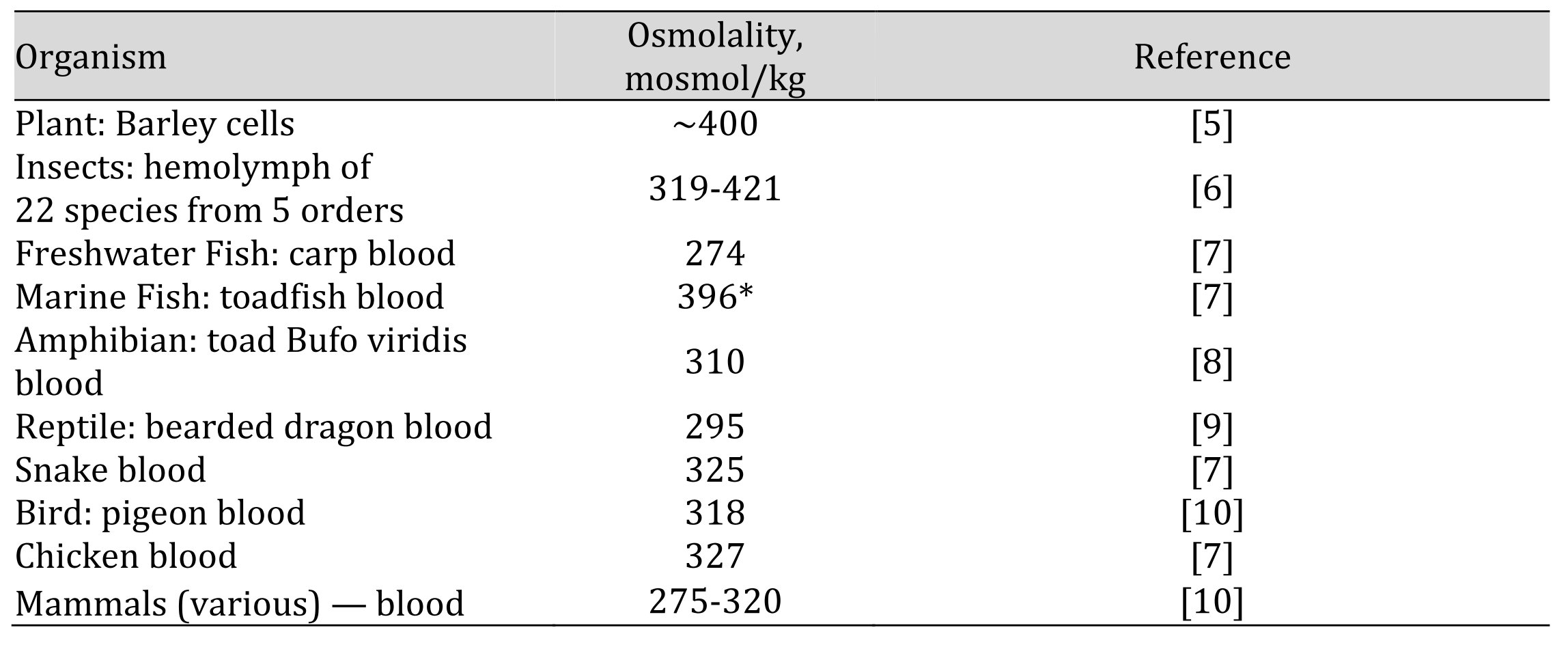
Table 1. Examples of internal osmolalities of selected aquatic and terrestrial osmoregulators. *approximately 40 mosmol/kg of this is due to TMAO
In short-term/transient volume disturbances, such as many animal epithelial cells routinely face, osmolytes in RVD/RVI are common inorganic ions (Na+, K+, Cl-), which are primarily regulated by channel, Na/K-ATPase pump and transport proteins in plasma membranes. These have been studied extensively by many researchers including Alexey Vereninov and colleagues (e.g., [15]) and many others (e.g., [1, 16]). Many cells, however, face long-term or permanent hyperosmotic stresses. Under such conditions, special intracellular organic osmolytes are typically used to regulate and/or maintain cell volume instead of inorganic ones. As has been thoroughly reviewed (e.g., [17-19]), these generally fall into a few chemical categories shown in Table 2, most of which have cytoprotective properties —such as protein stabilization—not found with inorganic osmolytes (see below). Because of these recent broad reviews, I will focus on one osmolyte, trimethylamine N-oxide (TMAO)— a solute in many marine animals that gives rise to 'rotting fish' odor arising from microbial breakdown into trimethylamine (TMA). It is a methylamine osmolyte which exhibits potent and possibly unique features for volume regulation and stabilization of macromolecules. Again because of other recent reviews [20-23], I will mainly discuss studies in the last few years along with selected key older ones.
How are these osmolytes used? As multicellular life arose, organisms evolved two different approaches to deal with elevated environmental osmolality:
1) Osmoconformers:
Most marine organisms from bacteria to animals—including some vertebrates (e.g., sharks, skates and hagfish)—are called osmoconformers: their body fluid osmolalities match that of the environment at ~1000 mosmol/kg in average seawater, and tracking higher or lower salinities (except at salinities below ~300 mosmol/kg). Their extracellular fluids (ECFs) are dominated by Na+ and Cl-, as in seawater; however, cells have only about 300–400 mosmol/kg from universal cell solutes. The remaining—about 600 mosmol/kg in full-strength seawater—is typically due to organic osmolytes:
Note that organic osmolytes are accumulated mainly inside cells, and much less so in ECFs (with a few exceptions such as urea in chondrichthyans; see below). As a recent example, in the deep-sea shrimp Benthesicymus cf. crenatus, TMAO in whole-muscle extracts was 241-272 but only 13-25 mosmol/kg in hemolymph [24].
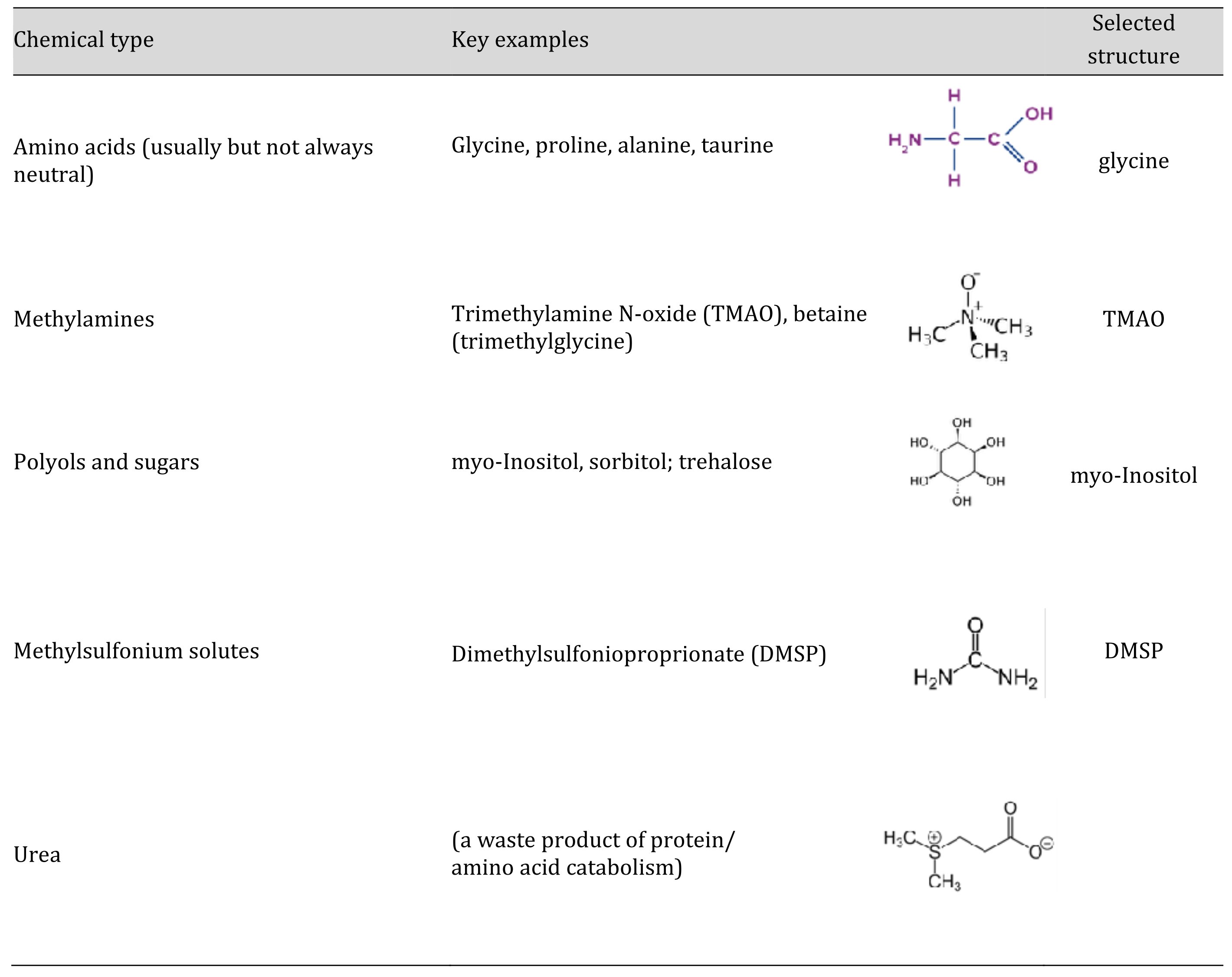
Table 2. Categories of organic osmolytes by chemical type with key examples and one selected structure for each
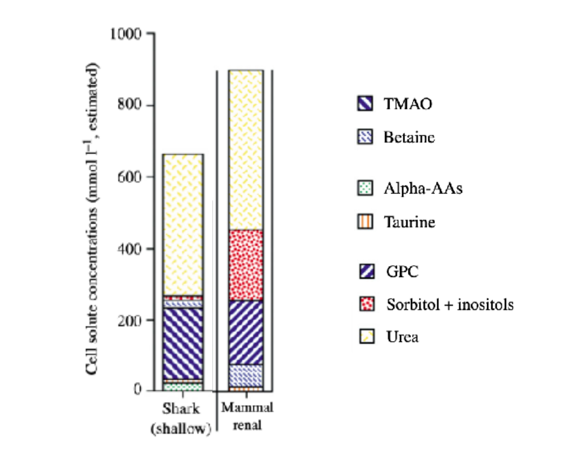
Figure 1. Cellular osmolyte content in sharks and mammalian renal inner medulla. Both osmoconform with urea, Na+ and Cl- in ECFs, and urea with methylamines (TMAO: trimethylamine oxide; GPC: glycerophosphorylcholine) and amino acids intracellularly, along with polyols in the mammal. Universal cell solutes, about 300-400 mosmol/kg, are not shown. Modified from [18].
2) Osmoregulators:
Most vertebrates, a few invertebrates such as estuarine crustaceans, and all freshwater organisms are called osmoregulators. These evolved specialized organs (e.g., gills and kidneys in bony fish) and behaviors (e.g., thirst and salt hunger) which maintain ECF osmolality at ~300- 400 mosmol/kg, so that individual cells are protected from volume changes (except transiently as in epithelial cells exposed to ingested fresh or saltwater). Organ-based osmoregulatory systems are found in all terrestrial animals; e.g., in mammals, plasma osmolality is maintained within a very narrow range (275 to 295 mosmol/kg in humans) primarily by kidneys and behaviors.
Osmoregulation in this sense was long thought to obviate the need for intracellular organic osmolytes. However, this is not the case. In mammals, osmoregulation usually does provide an osmotically stable environment for most cells, but there are major exceptions. In particular:
As noted earlier, organic osmolytes have protective properties not found in inorganic ones. The original and still widely used explanation for usage of organic osmolytes in long-term hyperosmotic stress is the 'compatibility' hypothesis [27], which states that these solutes—unlike inorganic ions—do not alter membrane potential nor perturb macromolecules' structures and functions. Indeed, most of them tend to enhance macromolecular stability, opposite to the effects of high Na+, K+, and Cl- concentrations on proteins [19] and DNA (breakage [28-29]).
Regarding organic osmolytes, the major exception to the compatibility hypothesis is urea, the primary osmolyte of chondrichthyan (as noted earlier). In addition to causing DNA damage in mammalian renal cells [30], urea is a well-known destabilizer of proteins through preferential binding to amino acids, which promotes protein unfolding (Fig. 2A).
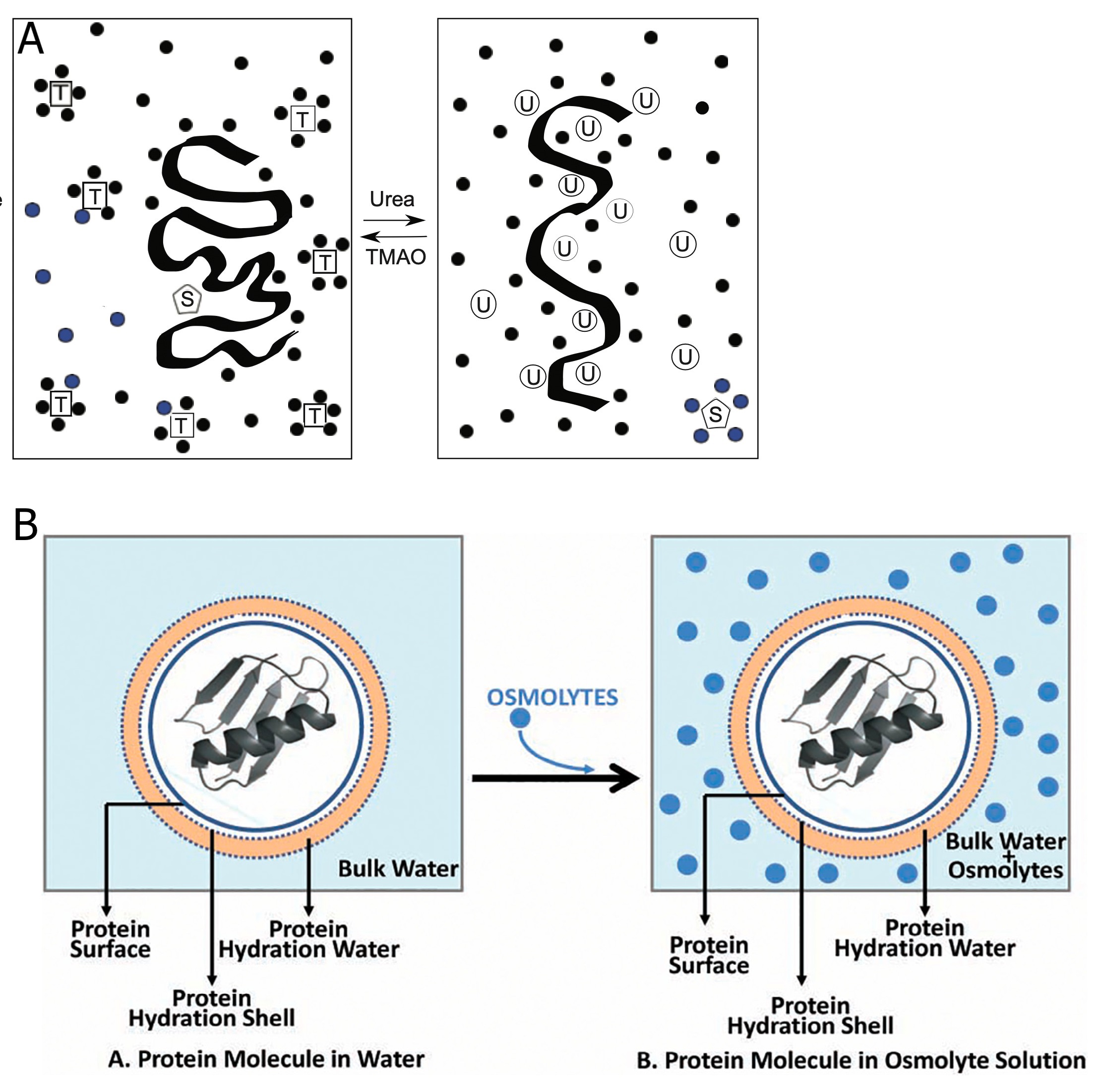
Figure 2. A. TMAO (T) and urea (U) with a protein (black strand) and substrate (S); small spheres represent water molecules. Right box: Urea binds to the peptide backbone and enhances unfolding and unbinding of substrate as that maximizes favorable binding sites. Left box: TMAO, excluded from the protein hydration layer presumably because of its own structured water layers (spheres around T), favors folding and substrate binding. Modified from Yancey and Siebenaller [19], with permission of The Company of Biologists. B. A schematic representation of a protein molecule in absence [left] and presence of preferentially-excluded stabilizing osmolytes [right], which do not enter and alter the protein hydration shell. From [31]: Sharma et al. (DOI: org/10.1016/j.ijbiomac.2021.02.102) under Elsevier License 5462140208130.
In contrast to urea, organic osmolytes that favor protein stability are typically hypothesized to work through preferential exclusion (or preferential hydration); i.e., they do not readily 'dissolve' in proteins' closest hydration layer and they reduce the availability of water for hydrating unfolding proteins (thus favoring folding; Fig. 2AB). Consequently, these solutes are called chemical chaperones by some researchers. Reduction of water availability arises, at least for some osmolytes, from strengthening of the water network; e.g., Meersman et al [32]., using isotopic substitution neutron-scattering measurements, found that TMAO oxygen strongly hydrogen-bonds to two to three water molecules, with the TMAO-water network being tighter than that of pure water. TMAO's methyl groups can interact with oxygen of water probably forming a type of clathrate structure (Fig. 3). TMAO and other stabilizers also greatly reduce several modes of water dynamics—e.g., translational and rotational—as well as water structure [34-35]. Due to effects on water, the stabilizers are also termed cosolvents or cosolutes [2, 36-38]. As Scherer [39] stated:
‘Protein interactions in water are also clearly mediated by the other solution components...Cosolutes, including the important biological osmolytes…are inextricably linked to...the stability and interactions of proteins in solution.
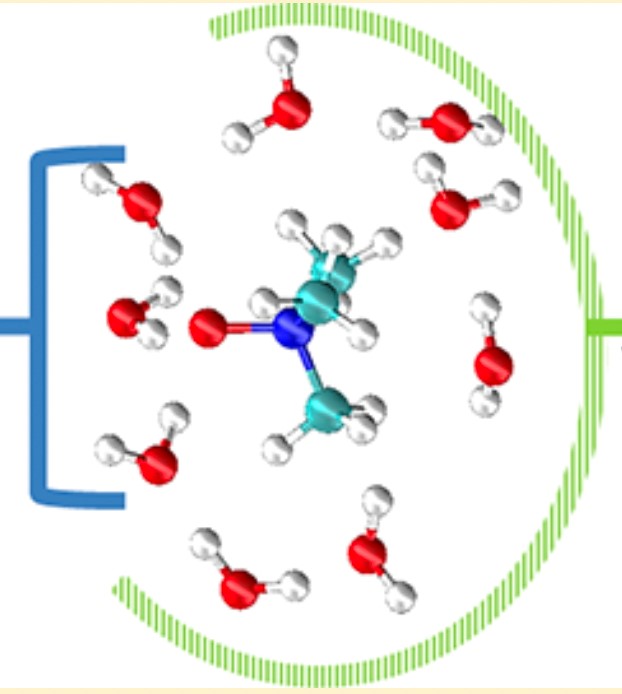
Figure 3. Model of water molecules around TMAO. Water molecules form hydrogen bonds with the oxygen (region indicated by the blue bracket), whereas they avoid the methyl groups building a clathrate-like cage around them (region indicated by the green curve). Reprinted with permission from [33]: Larini and Shea (DOI: org/10.1021/jp403635g), copyright 2013 American Chemical Society.
The water interactions have also been referred to as (micro)molecular crowding (e.g., [40]). It is not clear this is a useful term as it appears to differ from macromolecular crowding in that, again, water dynamics/structure are strongly affected by so-called (micro)molecular crowding agents like osmolytes but not by macromolecular crowding [41]. Regardless, these concepts are often presented in a broad way that implies most organic osmolytes are alike and interchangeable in this regard. This is misleading and possibly incorrect in two important ways.
First, organic osmolytes differ greatly in cytoprotective metabolic and chemical properties beyond protein stabilization and basic osmotic effects. Key examples are antioxidation (e.g., some polyols, DMSP, taurine), redox balancing (e.g., many polyols), sulfide detoxification (hypotaurine), and predator repellent (DMSP). However, this is a separate issue from general protein stabilization and water structure effects; it has been reviewed extensively elsewhere [18-19] and will not be discussed further here.
Second, and the main focus of this review, stabilization of proteins may not be attributable to identical preferential exclusion effects among all osmolytes. This is perhaps best exemplified by TMAO with its potent stabilizing/counteracting properties in at least two contexts in the oceans:
Counteracting urea: At 2:1 urea:TMAO (Fig. 1), as found in most chondrichthyans, TMAO's stabilizing abilities counteract urea's destabilizing effects on a variety of macromolecular processes. Other osmolytes can do so as well, but TMAO is often the most potent and may have unique mechanisms; see TMAO vs urea section below.
Counteracting hydrostatic pressure (HP): TMAO concentration increases linearly with depth in many marine animals (e.g., cnidarians, crustaceans, cephalopods, fishes), from about 40 mM in shallow bony fishes to ~400 mM in the the deepest fishes (the Kermadec and Mariana trench snailfishes at 7000 - 8000 m; Fig. 4A). In sharks and rays—chondrichthyans—an increase in TMAO with depth is offset with a decrease in urea, reversing the urea:TMAO ratio from 2:1 to 1:2 in species from ~3000 m (Fig. 4B). These trends correlate with depth limits proposed for bony (~8400 m) and chondrichthyan (~4000 m) fishes [42- 44]. In invertebrates, the increase in TMAO with depth is offset with a decrease in glycine and other osmolytes (Fig. 4C1, 4C2) [45-46]. Pressure is the only environmental factor that increases linearly with depth, and numerous tests have found that TMAO counteracts the destabilizing effects of pressure on protein structure and function better than other common osmolytes. It has been termed a piezolyte (pressure solute) for this property [47]. See hydrostatic-pressure section below.
In terms of molecular properties, TMAO often stands out as an osmolyte in several ways.
Volume Regulation. TMAO is a 'superosmolyte' in that is has the lowest self-solvation values (see Fig. 5 caption) and highest osmotic coefficient among common osmolytes. An ideal colligative solute has an osmotic coefficient of 1.00; a value >1.0 indicates a stronger ability to retain cell water against osmotic losses. Key osmotic coefficients at 1 M are shown in Table 3. Also TMAO has little effect on solution density; Table 3 shows how TMAO differs in this regard at 25˚C from other osmolytes and seawater [48-50]. At a temperature closer to the deep sea—5˚C—and physiological concentrations, density of water increases with solute concentration as follows [51-52]:
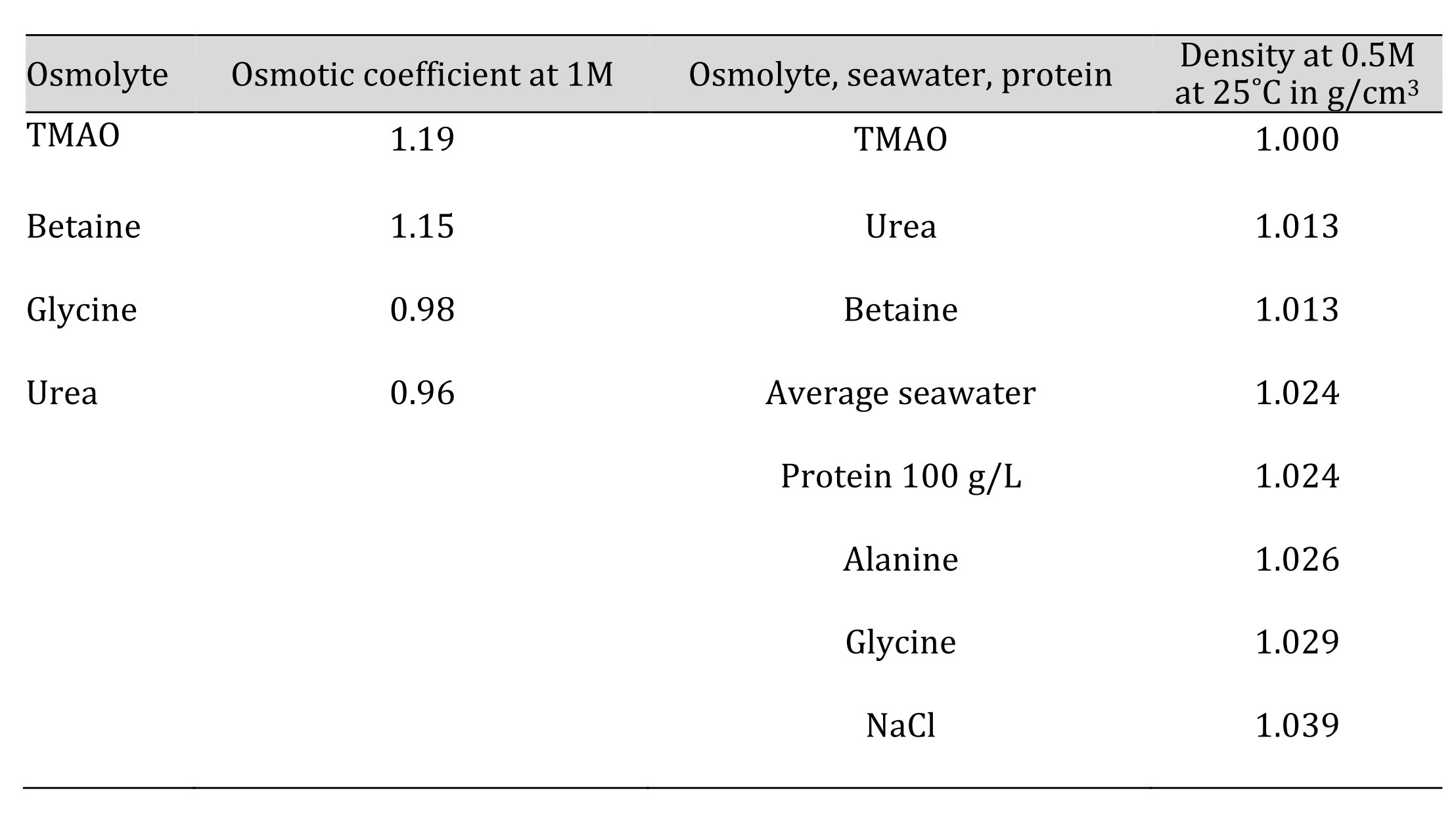
Table 3. Osmotic coefficients [48-49] and solutions densities in g/cm3 of 1 M solution at 25˚C (from [50])
These are at atmospheric pressure; see TMAO vs pressure section below for HP effects. In short, TMAO should retain cell water more effectively than most osmolyes while at the same time having no significant effect on cell density, which may be beneficial if density homeostasis is important to cellular processes [3].
Chaperoning proteins. TMAO's high osmotic coefficient reflects interactions with water (Fig. 3) that presumably contribute to its protein-stabilizing effects as well as cell volume regulation. In this regard, TMAO is the strongest in terms of the so-called 'osmophobic' effect [53], which refers to 'repulsion' of a generic protein backbone from an osmolyte solution, favoring protein folding under the preferential exclusion model. Shown in Fig. 6A is the ∆g of transfer of the peptide backbone to a 1M osmolyte solution [54]; a model of this osmophobic effect for TMAO is shown in Fig 6B [55]. Note that TMAO exhibits the strongest unfavorability for this transfer, with the trimethylamine betaine being second; in turn, the trimethylamines are stronger than other common osmolytes (Fig. 6A; see also [19] for a review). Denaturants show favorability.
Other studies have expanded on osmophobic mechanisms. Examples include these:
Chaperoning nucleic acids and membranes. Beyond proteins, TMAO has also been found to have stabilizing effect on other large molecular structures. For membranes, TMAO has been found to be excluded from the hydrated lipid membrane surface due to unfavorable interactions with polar lipid head groups, leading to dehydration of the membrane and an increase in phospholipid packing density. TMAO also raises the fluid/gel phase transition temperature [59-60] (Fig. 7). For nucleic acids, TMAO stabilizes tertiary structures of both DNA and RNA, due in part to exclusion from and dehydration of the phosphate backbone [61-63].
Chaperoning liquid−liquid phase separation (LLPS). LLPS is perhaps the latest biological phenomenon to be tested with TMAO. This is a broad term for the process of macromolecules and molecular complexes with similar properties forming droplets or condensates in cells without needing membrane boundaries. These membraneless 'pseudo-organelles' include nucleoli and ribonucleoprotein (RNP) 'stress granules' for mRNA storage, processing and translational regulation, which may form and dissipate rapidly under varying stress conditions. In some cases, condensates can be pathological fibrils. TMAO greatly affects LLPS; for example, Choi et al. [64] found that TMAO enhances the beneficial form of RNP condensation while inhibiting harmful fibrillar aggregation.
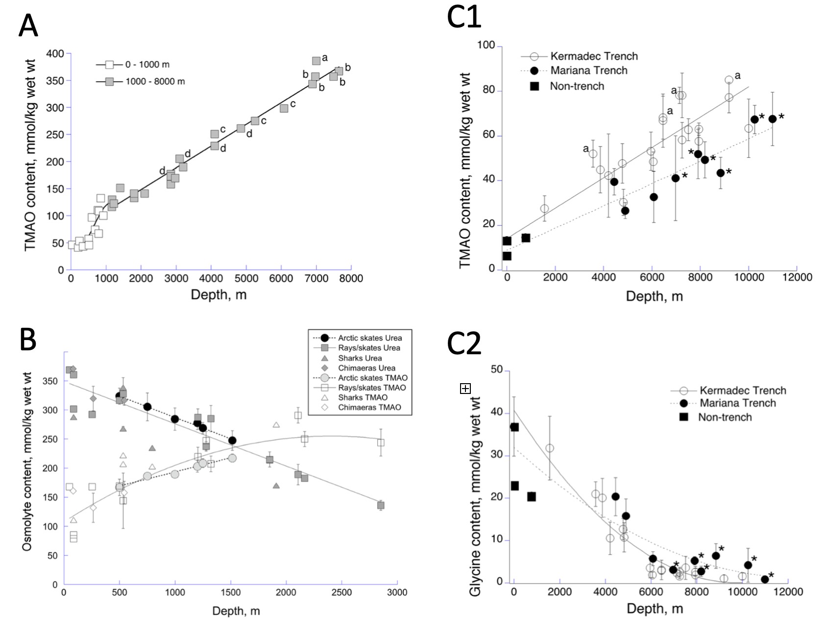
Figure 4. TMAO (mmol/kg) in muscle tissue vs depth in the ocean. A) Osteichthyans (bony fish); aKermadec Trench hadal snailfish Notoliparis kermadecensis; bMariana Trench snailfish Pseudoliparis swirei; cgrenadier Coryphaenoides yaquinae; dAbyssal grenadier Coryphaenoides armatus. Modified from Yancey et al. [42], Linley et al. [43] and Yancey [20]. These fish would become hyperosmotic below ~8200-8400 m, possibly preventing them from living at greater depths (see [42]). B) Chondrichthyans (cartilaginous fish), which may also have depth limits due to osmolytes; adapted from Laxson et al. [44], Yancey et al. [45] and Yancey [20]. C) Amphipods; modified from Downing et al. [46] and Yancey [20].
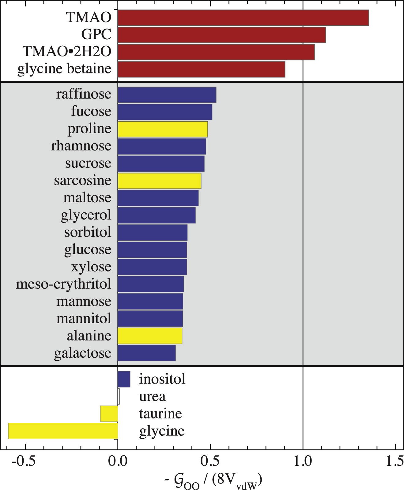
Figure 5. Osmolyte self-solvation values G00 normalized to -8VvdW , the hard-sphere limit extrapolated to 0 M concentration (VvdW is a solute's van der Waals volume). Self-solvation refers to a solute associating with identical solutes in solution. A value around 1.0 for -G00 / 8VvdW (abscissa) indicates solutes with large negative self-association values, acting like hard spheres incapable of overlapping/associating with each other (i.e., little or no self-solvation). The osmolytes fall into three groups, indicated by gray or white backgrounds. All trimethylamines are in the top group (in red) exhibiting nearly ideal hard-sphere behavior, presumably also explaining exclusion from peptide backbones [49]. In the bottom two groups, carbohydrates are indicated in blue, and amino acids in yellow; the bottom molecules exhibit the most self-association (i.e., self-solvation). From [49]: Jackson-Atogi et al. (2013) (DOI: org/10.1016/j.bpj.2013.09.019) under the Creative Commons CC-BY-NC-ND license.
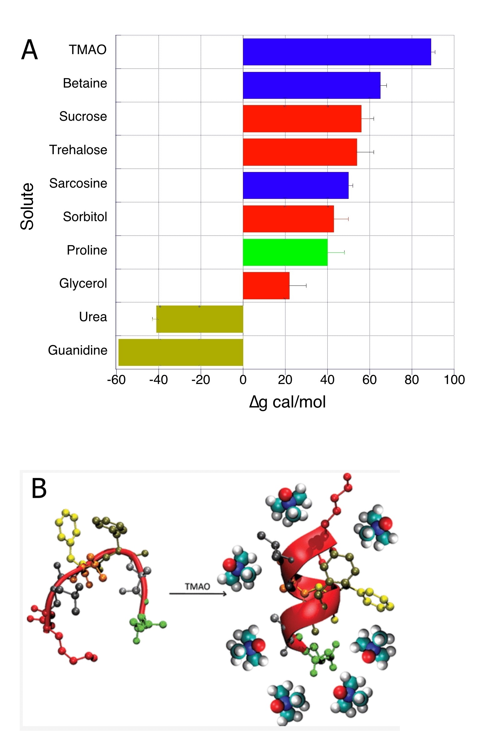
Figure 6. A: Thermodynamic Δg (cal mol−1) of transfer of peptide backbone to 1 mol l−1 of indicated osmolyte, plotted from data in Street et al. [54]. Dark yellow = denaturants; red = carbohydrates; green = amino acid; blue = methylamines. Note TMAO’s positive (unfavorable) Δg at +89 is about double the favorable negative Δg for urea −41; i.e., TMAO’s folding effects are twice as effective as urea’s unfolding effects, thermodynamically canceling at about 2:1. B: Model of TMAO's osmophobic effect, here favoring alpha-helix folding with exclusion from protein hydration layer. Reprinted with permission from [55]: Cho SS, Reddy G, Straub JE, Thirumalai D; J Phys Chem B. 2011;115:13401-13407; copyright 2011 American Chemical Society.
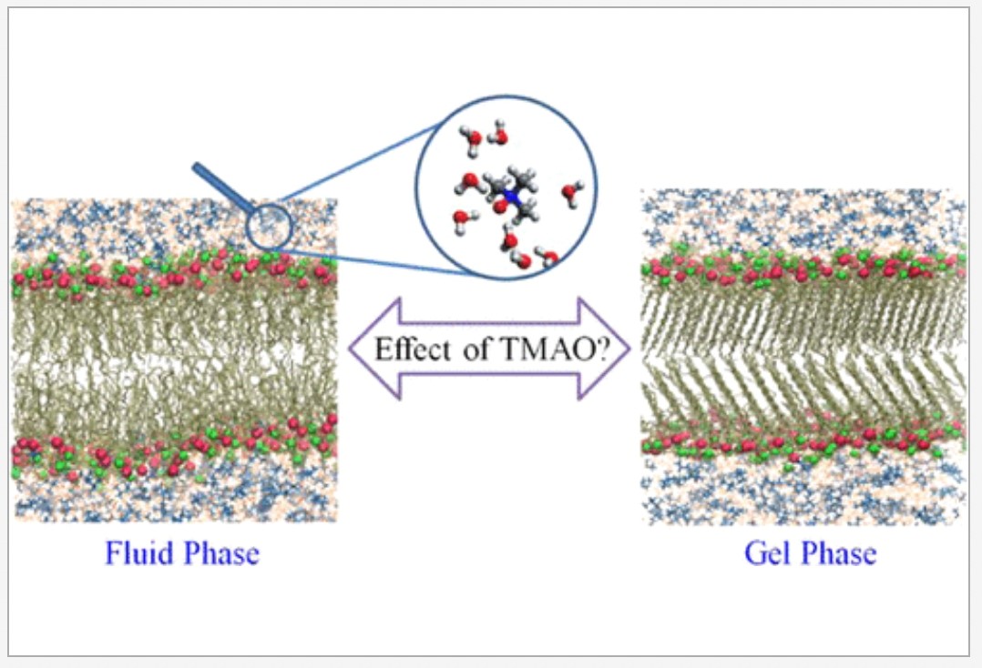
Figure 7. Model of TMAO's interaction with fluid and gel phase of phospholipid bilayer. Reprinted with permission from [60]: Maiti A, Daschakraborty S. J Phys Chem B 2021;125:1167−1180. Copyright 2021 American Chemical Society.
As noted earlier, TMAO is often the most potent stabilizing osmolyte in nature; but the question remains: is this a matter of degree or due to some unique mechanisms? Many studies conclude the former (e.g., [65]), i.e., a universal exclusion mechanism, but others do not. Such studies often involve comparing TMAO with the common trimethylamine osmolyte betaine (or glycine betaine) and with key non-methylamine solutes. An illuminating example of the latter is glycine, an osmolyte widely found in marine invertebrates in the absence of any apparent protein perturbants, but not at high levels in urea-rich or deep-sea cells (Fig. 4C2). TMAO and glycine have the same molecular weight (75 daltons), but TMAO is far more compact with a much smaller charge separation and a trimethyl component (Table 2), which should create important differences including close charge separation/dipole moment and clathrate water structure (Fig. 3). Laurent et al. [66] (see Pressure section later) concluded:
'Protein surfaces, by definition, must be more hydrophilic than their hydrophobic core. Osmolytes with large hydrophobic character are therefore more likely to be excluded from the protein surface and promote biomolecular stability. We therefore propose that stabilising osmolytes require both large hydrophobic character to promote stabilisation of the surrounding water network, and preferential exclusion and hydrophilic character to promote stabilisation of the surrounding water network and allow them to be sufficiently soluble. In this respect, TMAO is ideal.'
In this regard, TMAO has indeed been found to differ significantly in effects on proteins and water compared to glycine (which lacks large hydrophobic characteristics), and betaine in some studies. See Sharma et al. [31] for a recent review on TMAO vs non-methylamine osmolytes. Illuminating and sometimes apparently contradictory studies on proteins include the following:
Glycine's properties often fit the 'compatibility' concept, but does TMAO? There are two problems here. First, TMAO's potency could make a difference in its usage in nature compared to less powerful stabilizers even if the mechanism is much the same. As noted earlier, unlike glycine and many other organic osmolytes, TMAO does not appear to used by nature at high concentrations (>50 mM) in the absence of a destabilizer of macromolecules—especially urea and high pressure [19-21, 75], and possibly intracellular NaCl [18]. This may be due to TMAO's potent effects creating a potential 'yin and yang' problem [76]: the strong stabilizing ability of TMAO can be very useful, but in the absence of a perturbing agent it can inhibit protein flexibility and favor nonfunctional protein aggregates; e.g., TMAO can favor prion and damaging amyloid formations [77] (see [19] for other examples), and TMAO switches from a stabilizer to a destabilizer for the protein stem bromelain as concentration increases [78]. In more recent examples, Bhat et al [79-80]. found that TMAO, but not betaine or sarcosine, favors non-functional aggregates of the intrinsically disordered α-casein protein—a process that urea counteracts in a reversal of the usual TMAO-urea counteraction story (see TMAO — urea section below).
As noted earlier, over-stabilization should disqualify TMAO and perhaps some other osmolytes from being considered 'compatible'. The complexity of stabilizing properties led Gilles [81] to propose the term 'compensatory' rather than 'compatible' solutes, though this term has not been widely adopted.
In conclusion regarding TMAO vs other chemical chaperones, it does appear that TMAO is unique in many ways, with some properties consistent with universal preferential exclusion but with some that add to (or conflict with) that broadly stated mechanism. However, how much those latter features contribute to or detract from its potent stabilization abilities, and how many features arise in simulations that need refinement, remain unclear. Thus, different methodologies, as well as different model systems, have not yet produced one consistent picture.
The story is quite different with urea, used as an osmolyte in nature but with dramatically opposite ('anti-chaperoning') effects on macromolecules. Since the discovery of TMAO-urea counteraction in the 1970s noted earlier (see [17]), numerous studies have been conducted on mechanisms. Again, there are some apparently inconsistent results. Most studies indicate that the two solutes act independently by preferential exclusion (TMAO through strengthening water structure and hard-sphere properties) and preferential binding (urea binding to peptide backbones with little effect on water structure) [82]. Thus the counteraction is often attributed to simple additivity of opposing effects without TMAO-urea interactions being important [83-84]. Notably, in Fig. 6A, urea has a favorable value for peptide unfolding ∆g that is about one-half the unfavorable value of TMAO's, providing a thermodynamic explanation for the 2:1 urea:TMAO ratio found in sharks and their relatives. In a recent review, Gao et al. [2] note that a 2:1 mixture of urea:TMAO shows almost ideal colligative behavior with additive cancelation in terms of osmotic coefficients, concluding:
'net repulsive or attractive interactions between TMAO and urea are lacking, which is a further evidence for a water-mediated mechanism of the counteraction.'
However, studies which contradict the independent-action model have found close TMAO-urea interactions. Key examples are as follows:
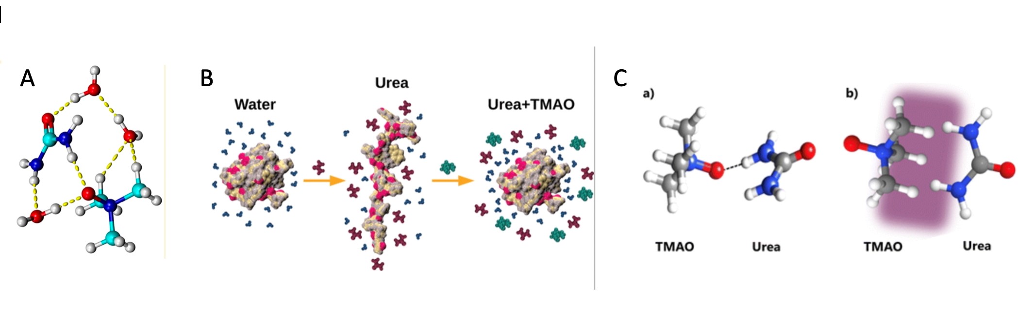
Figure 8. Possible Urea and TMAO interactions. A) Proposed urea-water-TMAO network, reprinted with permission from [86]: Zetterholm et al. (DOI: 10.1021/acs.jpcb.8b04388). Copyright 2018 American Chemical Society. B) Model showing urea binds to the peptide backbone, favoring unfolding; TMAO is preferentially excluded, but may also interact with urea, reducing its penetration into the peptide. From [88]: Ganguly et al. 2020 (DOI: 10.1021/acs.jpcb.0c04357) under EMBL-European Bioinformatics Institute Open Science. C) Two possible conformations of a TMAO–urea complex: (a) is hydrogen bonded (black line), and (b) is hydrophobic interaction in the purple zone. Carbon – grey, hydrogen – white, nitrogen – blue, and oxygen – red. The authors' data were most consistent with (a). From [89]: Nasralla et al. 2022 (DOI: .org/10.1039/D2CP02475F) with permission under Creative Commons Attribution 3.0 Unported Licence.
This somewhat confusing array of findings reveal possible contradictions, but as noted earlier, in light of the many different methods used—including simulations rather than pure experimental data—the mechanisms proposed are not necessarily in conflict nor mutually exclusive. More work is needed to sort out whether direct TMAO-macromolecule, TMAO-urea interactions, and other non-exclusion mechanisms—even if real—are important in enhancement or inhibition of macromolecular structure and function. The following sections return to that issue.
Counteraction with proteins. Even within the independent-action model, some studies suggest effects that complicate the basic preferential exclusion model—recall the surfactant hypothesis earlier [71]. More recently, Su and Dias [90], using MD and model peptides, found that TMAO favors the exposure of non-polar side chains to water, questioning the simple exclusion mechanism; but they also found that TMAO increases the magnitude of proteins' charge-charge interactions favoring intra-backbone interactions and protein folding, in accordance with exclusion. Overall, the folding forces dominated with their model peptide. Urea, in line with previous studies, independently destabilized hydrophobic and intra-backbone interactions.
Counteraction with nucleic acids. TMAO and urea have opposite effects on nucleic acids which again appear to be independent. Holmstrom et al. [63] conclude from a study on RNA and DNA structures that:
'TMAO always stabilizes nucleic acid structure formation by increasing the folding rate constant and decreasing the unfolding rate constant… Conversely, urea always destabilizes nucleic acid structure formation by decreasing the folding rate constant and increasing the unfolding rate constant.'
Recent examples include the following:
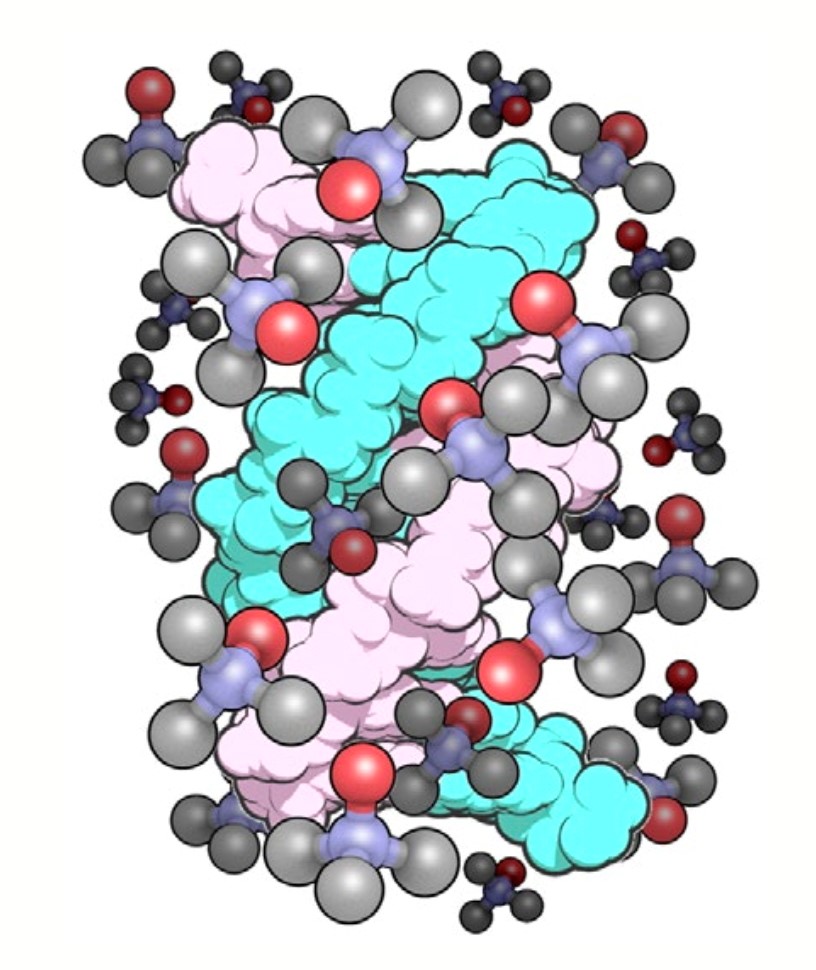
Figure 9. Possible DNA-TMAO interactions, with TMAO binding to the groove of the classic DNA duplex. TMAO molecules are shown as red oxygen, blue nitrogen and grey methyl groups. From [94]: Ueda et al. 2016 under Open Access Creative Commons Attribution License (http://creativecommons.org/licenses/by/4.0/)Possible DNA-TMAO interactions, with TMAO binding to the groove of the classic DNA duplex. TMAO molecules are shown as red oxygen, blue nitrogen and grey methyl groups. From [94]: Ueda et al. 2016 under Open Access Creative Commons Attribution License (http://creativecommons.org/licenses/by/4.0/)
Counteraction with membranes and lipids. For membrane lipids, counteraction also occurs with urea destabilizing and TMAO stabilizing lipid bilayers (e.g., Valerio et al. [95]), but this can be rather complex, as found in three recent studies:
Returning to the primary question of this review—is TMAO unique as a protein stabilizer? - it is important to note that other chaperoning osmolytes may be used to counteract urea in nature (at least for proteins, which have received far more study than nucleic acids and membranes).
Clearly there is much to learn about chemical chaperones' stabilizing mechanisms, with preferential exclusion currently remaining as the primary unifying mechanism but with effects on water structure, synergistic interactions in mixtures, and direct interactions with macromolecules yet to be fully understood. For example, while TMAO is unique in its compact structure, the large GPC molecule works as well in some cases but its mechanisms are unknown. To explore this issue further, there is considerable evidence of TMAO's uniqueness in a very different context—that of high hydrostatic pressure in the deep sea.
The most recent discovery of TMAO in nature—increasing concentrations with depth in many marine taxa (Fig. 3) in parallel with increasing hydrostatic pressure (HP) [109]—has yielded another chaperoning hypothesis for TMAO, namely as a piezolyte. HP, increasing 1 atm or ~0.1 MPa per 10 m in lakes and seas, ranges from 1 atm (0.1 MPa) up to 1100 atm (111 MPa) in the Mariana Trench at 11 km deep; pressures above roughly 50-100 atm (5 - 11 MPa) have significant inhibitory effects on proteins, nucleic acids and membranes. Specifically, HP inhibits processes with positive volume changes—e.g., many cases of macromolecular folding and ligand binding—by trapping dense water around charged moieties, compressing proteins, and, at higher levels, pushing water into proteins' interior voids (Fig. 10) [110]). As Roche and Royer [111] express this:
'…the main contributing factor to the decrease in volume between the folded and unfolded states of proteins is the existence in folded protein structures of solvent excluded void volumes that are eliminated upon unfolding'.
High HP can also dissociate transcription factors from DNA and protein subunits from each other, but for some proteins it can trigger the formation of non-functional aggregates [110]. How does life survive all this? Certainly, evolution of pressure-resistant structures has occurred, but pressure-counteracting piezolytes like TMAO may be of equal importance.
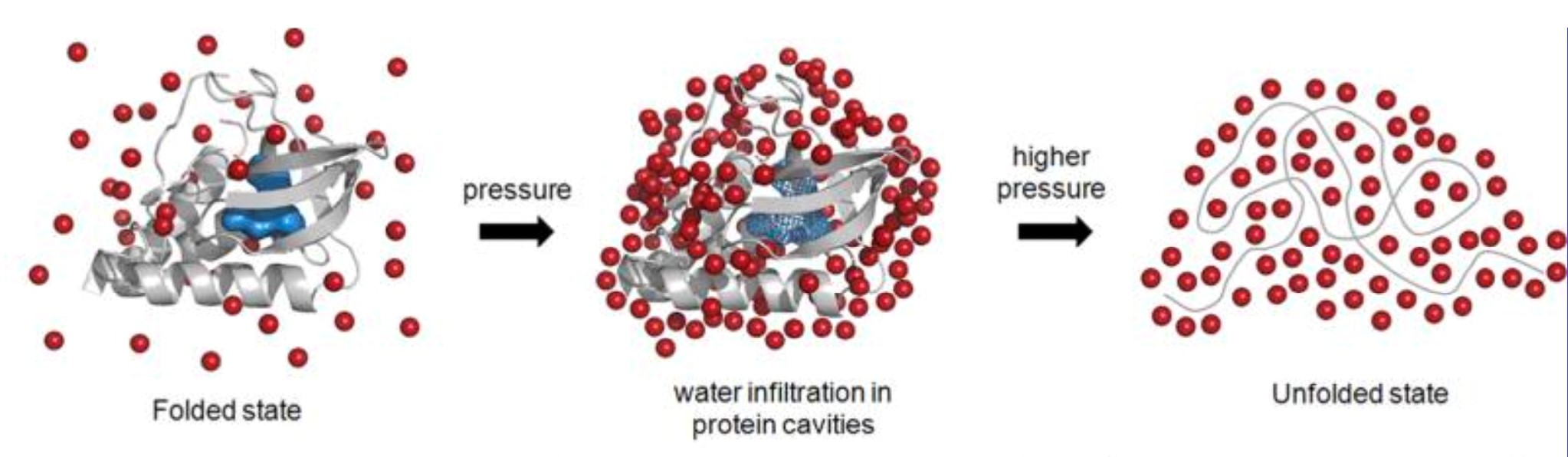
Figure 10. Schematic representation showing HP effect in water-excluded cavities (blue surface within the protein model). A staphylococcal nuclease was used for illustration. Under pressure, water molecules (red spheres) infiltrate into the protein leading to cavity disassembly (blue mesh within the protein model) and unfolding. From [110]: Silva et al., https://pubs.acs.org/doi/10.1021/cr400204z; further permissions related to the material excerpted should be directed to the American Chemical Society.
Counteraction with proteins. Regarding deep‐sea proteins, some have evolved amino acid substitutions (most poorly understood) that reduce HP sensitivity, e.g., larger branched side-chains that reduce voids inside proteins [112]. However, many (perhaps most) deep-sea proteins are incompletely adapted to high HP. Many such proteins have been found to be 'rescued' at high HP by TMAO [19-20, 113] (Fig. 11). How does TMAO do this counteraction? Numerous studies have shown that TMAO's strengthening of the water network prevents high HP's effects on water interactions with proteins and charged reactants. As at low HP, TMAO enhances water hydrogen bonding, but a different feature arises at high HP: TMAO is excluded from a protein's hydration shell, but accumulates just outside this first shell in the 2nd hydration shell which TMAO then 'pushes' outwards in opposition to compression by high HP [114]. This prevents high HP from pushing water into protein voids [115-116]. It can also prevent some proteins from contacting each other at high HP, thus preventing some non-functional aggregates [117].
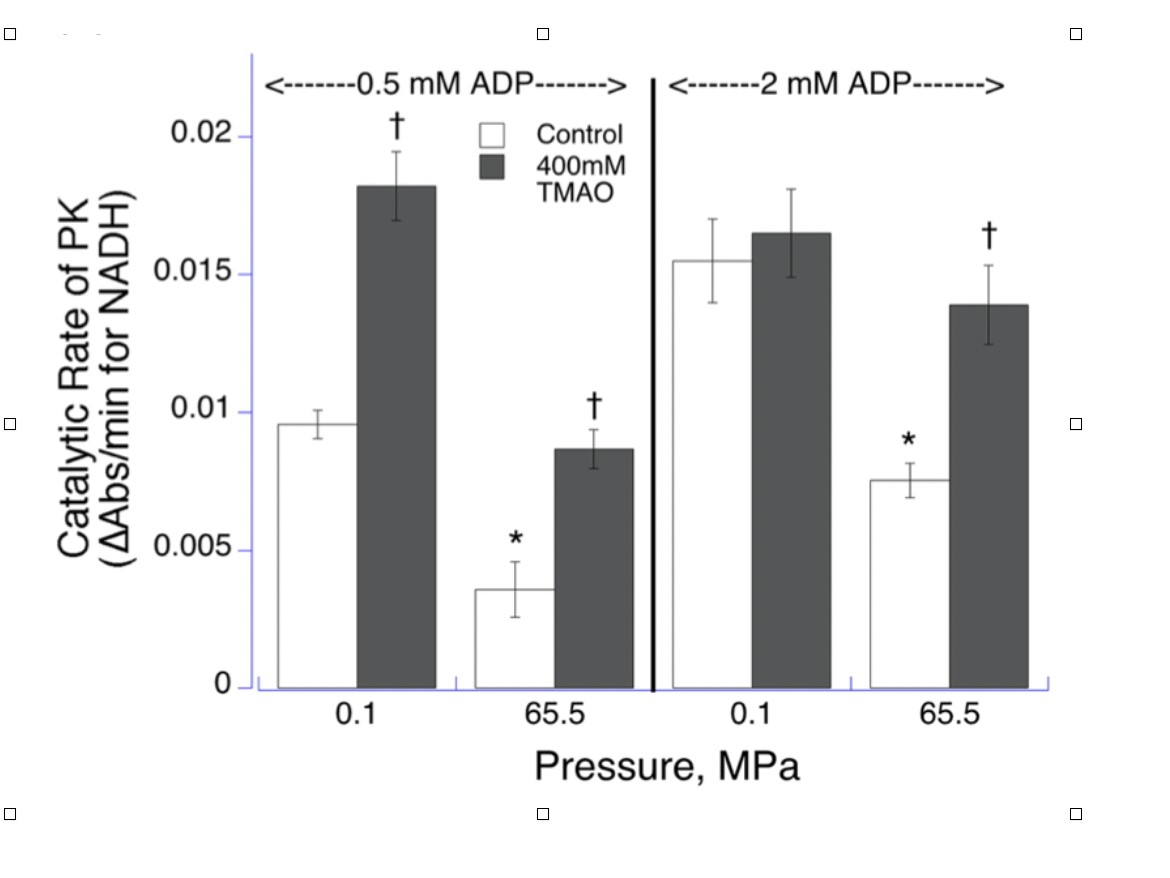
Figure 11. Catalytic rate of pyruvate kinase (PK) from Notoliparis kermadecensis (Kermadec Trench snailfish from 7,000 m) at low and high hydrostatic pressure, for two concentrations of ADP at 5°C. *Significant inhibition compared with 0.1 MPa control; †significant enhancement by TMAO over controls at same pressure. From [113]: Gerringer et al. 2017 under Open Access Creative Commons Attribution License (http://creativecommons.org/licenses/by/4.0/).
All this appears to fall under the preferential exclusion model (Fig. 12) [118]; moreover, TMAO is not unique as a piezolyte in laboratory testing, and possibly not in nature. Other solutes can counteract HP effects, for example, on tubulin assembly, which pressure destabilizes and both TMAO and sucrose stabilize. However, sucrose requires a much higher concentration to do so [119] and is not a piezolyte in nature. In the oceans, some animal taxa do not have TMAO (e.g., echinoderms), while others have TMAO but also other solutes that increase with depth, e.g., amphipods, in which TMAO dominates but in which other potential piezolytes—notably GPC and scyllo-inositol—increase with depth, while betaine and glycine (Fig. 4C) decrease. TMAO ranks highest as a piezolyte among common osmolytes in many studies [118, 120-121], so the possible role of these other solutes in depth adaptation is uncertain. Notably, betaine increases with depth in some animals, but only to moderate depths [120, 122]. The other potential piezolytes (e.g., GPC, scyllo-inositol) may have properties under the highest pressures in nature that TMAO lacks [46]; see Membranes section below. All of this hints at unique features of some stabilizing osmolytes. For TMAO itself, recent findings suggest unique features, many of which arise only under high HP:
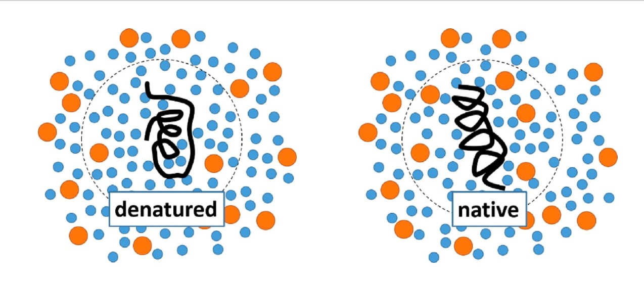
Figure 12. A pressure‐denatured protein (left) has water (blue) penetrating its interior and a smaller volume than the native state. TMAO (orange) binding of water favors the native state (right). Note that TMAO is excluded from the immediate hydration layer of the protein. From Shimizu and Smith [118] with permission from RightsLink license 4758350992662.
Counteraction with nucleic acids. Though less studied, TMAO can counteract effects of high HP on nucleic acids. While the classic DNA duplex is not very pressure sensitive, other form such as the telomeric G-DNA quadraplex (G4) and DNA/RNA hairpin turns are. In one study, Knop et al. [127], using Förster resonance energy transfer (FRET), found that TMAO and macromolecular crowding agents 'are able to effectively rescue the G-DNA quadraplex from unfolding in the whole pressure range encountered on Earth'. TMAO has a great advantage in nature over crowding agents because it does not alter cell density. Similar results using FRET have been reported for a DNA hairpin [128]; for G4 DNA, DNA hairpin, and RNA 'thermometer' hairpin structures [129], and for the lysine riboswitch gene-expression regulator [130].
Counteraction with membranes and lipids. The story is more complex for membranes, because TMAO's stabilizing effects on membranes seems likely to exacerbate the compacting of membranes under high HP, rather than counteracting it. One possibility is that the other potential piezolytes found in some animals (e.g., amphipods; [46]) have stabilizing properties with membranes under pressure, but this remains to be determined. See Somero [21] for a recent analysis.
Solution Properties and Volume Regulation. In addition to effects on macromolecules, high HP also compresses water itself and thus cell volume, up to about 5% at 11 km, with potential negative effects through increased crowding / cell density. However, TMAO uniquely does not alter cell density (Table 3) and makes water largely incompressible (see below), reducing cell volume compression and density increases (hypothesized by Somero et al. [131]. Recent studies on TMAO solutions under pressure reveal aspects that again suggest that TMAO is unique:
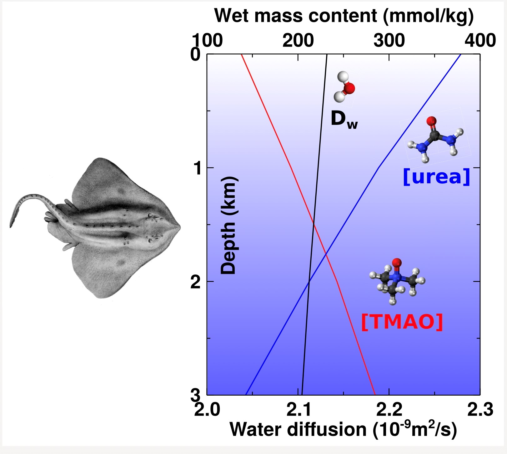
Figure 13. Diffusion rate for water (bottom axis) vs depth in the presence of urea (blue line), TMAO (red line), and urea:TMAO (dotted line) at physiological contents of these osmolytes (top axis) found in chondrichthyans (see Fig. 4B). Reprinted with permission from [132]: Teng X, Ichiye T. J Phys Chem B 2020;124:1978−1986. Copyright 2020 American Chemical Society.
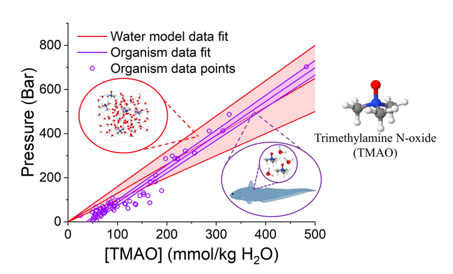
Figure 14. Agreement between model system and whole animals. The pressure-resisting ability of TMAO as calculated by considering the perturbation to water − water hydrogen bonding by TMAO addition and pressure (red) through neutron scattering. This is compared with data reported by Yancey et al. [42] (purple) on the muscle TMAO contents of bony fish and the pressure of collection depth. From Laurent et al. [66] under Open Access Creative Commons CC BY license.
Counteraction with membranes and lipids. Counteraction in liquid−liquid phase separation (LLPS). In some cases, these important biological condensates/droplets (described earlier) can be very pressure-sensitive, with increase HP often causing dissipation of the droplets. TMAO has been found to effectively counteract this pressure effect for several condensates [134-135] including post-synaptic densities important in synaptic transmission (Fig. 15) [136].
TMAO Biosynthesis. A final question just recently being addressed is the source of TMAO in the deep sea. Enzymatic synthesis and diet are typical sources, with new details emerging:
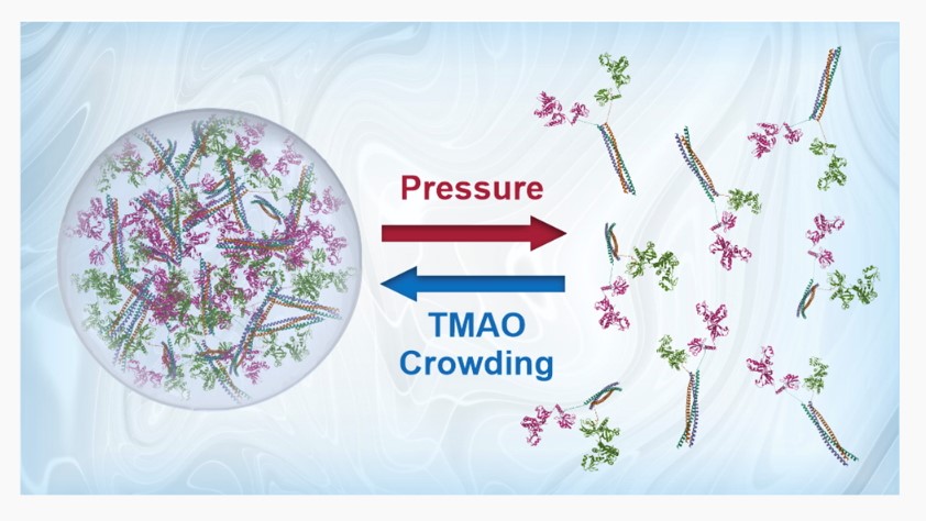
Figure 15. Model of cellular condensate formation via Liquid−liquid phase separation (LLPS), showing pressure disruption and TMAO stabilization. Reprinted with permission from [136]: Cinar H, Olivia R, Wu H, Zhang M, Chan HS, Winter R. J Phys Chem B 2022;126:1734–1741; Copyright 2022 American Chemical Society.
TMAO clearly exhibits many unusual and possibly unique properties compared to other common osmolytes / chemical chaperones. More research is obviously needed, and not for just pure academic interests. As reviewed elsewhere [18-21, 75] stabilizing osmolytes have many practical applications in medicine, agriculture, biochemistry, and in inspiring the design of new useful chemicals. Surprisingly, numerous plants (Arabidopsis, tomato, maize, etc.) have recently been found to make TMAO with flavin monooxygenases in response to various stresses including low water and salt stress. TMAO enhances survival and exhibits chaperone activity with plant proteins, suggesting applications for improving crop stress tolerance [142].
In medicine in particular, TMAO in humans is a controversial topic. On one hand, there are many proposals to use TMAO therapeutically to rescue malformed proteins in so-called protein conformational diseases (PCDs); e.g., TMAO restores binding of a non-functional mutant glucocorticoid receptor to glucocorticoids in chaperone complexes [143]. On the other hand, as discussed earlier regarding over-stabilization, some of those malformed proteins are favored by TMAO under some circumstances; e.g., TMAO promotes aggregation of the amyloidogenic intrinsically disordered peptide Aβ42 [144]. These are reviewed by Kumari et al. [145]. who document that 24 of 27 PCDs are rescued by TMAO in vitro (e.g., cystic fibrosis, Huntington's and glaucoma), but the rest (Alzheimer's amyloid, α-Synuclein, prions) are exacerbated. Moreover, elevated plasma levels of TMAO, generated by gut microbes from carnitine in beef, have been found to correlate strongly with atherosclerotic cardiovascular and possibly other diseases [146]. However, causation is not clear, with other studies showing cardioprotective features of TMAO in humans [147] and implicating TMA instead as the harmful agent [148-149]. Given the 'yin and yang' nature of this powerful stabilizer, perhaps it is not surprising that conflicting results are reported. Research on TMAO will continue, but more studies are also needed on other common osmolytes and on possible synergistic interactions in osmolyte mixtures also common in nature.
The author wrote the entire manuscript. The author thanks George Somero for informative email discussions and sending relevant publications regarding key topics in this review.
Statement of Ethics
The author has no ethical conflicts to disclose.
The author has no conflicts of interest to declare.